


 86 / 96

F
alcão
and
C
ampos
1096
R
ev
A
ssoc
M
ed
B
ras
2017; 63(12):1090-1099
of the modulation of the inflammatory profile was per-
formed according to Falcão et al.
52
The receptors were iden-
tified by flow cytometry, and their fluorescent histograms
were prepared so that the absolute number of cell surface
receptors labeled with the anti-receptor fluorescent mono-
clonal antibody of interest was generated, or the mRNAs
for genes of the receptors were detected. As a result, they
found an increase in the production of T
regs,
CD4
+
HLA-DR
+
,
a decrease inCD8
+
CTLA-4
+
and an increase in the expres-
sion of IL-10 by T
regs
up to the fourth week, with a mean of
the absolute number of receptors maintained after the sixth
week. The expression of CD8
+
INF-
γ
+
and CD14
+
INF-
γ
+
was
markedly decreased in the samples evaluated.
52
Thus, it is conclusive that the monitoring and ma-
nipulation of proinflammatory interleukins has the po-
tential to assist in the prognosis of anti and pro-inflam-
matory and degenerative changes in situ, monitoring the
course of the disease.
S
ynthesis
of
tracers
for
in
vivo monitoring
Image monitoring of symptoms of autoimmune diseases,
such as RA, is preferable considering that such a technique
will directly contribute to the accuracy of the diagnosis
and consequently the establishment of the therapeutic
mode and its intensity.
57
The accurate definition of the
site with a design of the inflammatory focus is relevant
in the choice of therapeutic management in RA.
58
Radio-
logical imaging, radiography, computed tomography,
nuclear magnetic resonance or ultrasound may favor an
analysis of the deleterious effects on the anatomical struc-
tures in the peripheral joints.
59
However, such images do
not aid in the early analysis of RA. Scintigraphy, on the
Recrutamento de
STAT
Formação do Complexo
STAT
com transloção
nuclear e ativação do
gene
B
A
M
MEMBRANA
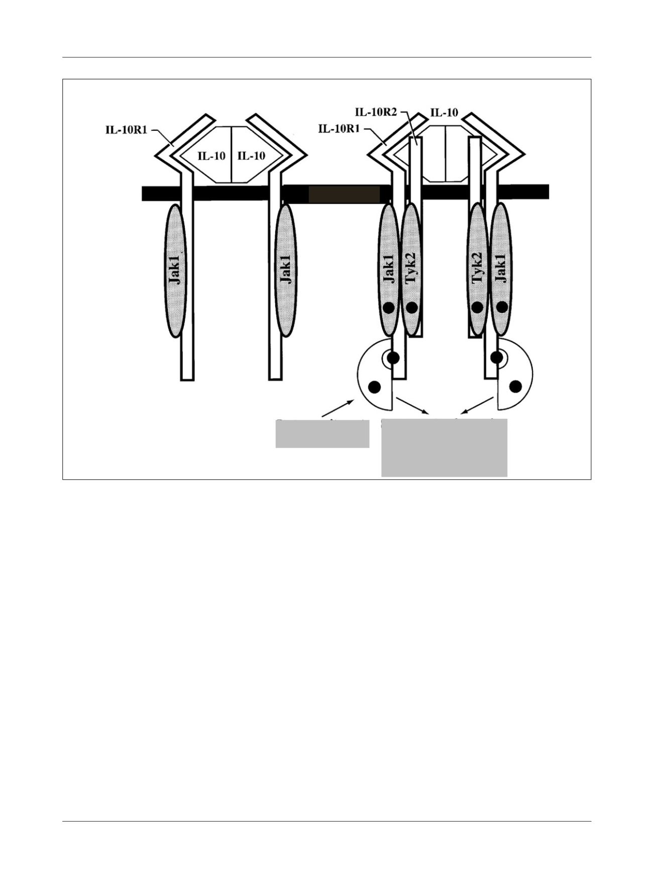
FIGURE 2
IL-10 receptor binding via
STAT-3
. A. Binding of IL-10 to IL-10R1 receptor via receptor-anchored
Jak-1
kinase. B. Binding of IL-10 to
IL-10R2 receptor, recruitment of
STAT-3
and
STAT
complex formation and gene activation by
Jak-1
and
Tyk-2
kinases. Both “outside-in” and
“inside-out” signaling are associated with distinct conformational changes in the extracellular segment. These changes vary with the type and
nature of the ligand and are modulated by divalent cations
.
(Adapted from Abbas and Lichtman,
1
2005.)
MEMBR E
STAT Recruitment
STAT complex formation with
nuclear translocation and
gene activation