


 23 / 116

G
iant
sclerosing
papilloma
mimicking
locally
advanced
breast
carcinoma
R
ev
A
ssoc
M
ed
B
ras
2014; 60(6):518-519
519
The core biopsy showed a benign complex papillary le-
sion. Since the radiologic and pathologic divergence did not
allow a definitive diagnosis ofmalignancy, an ultrasound-gui-
ded stereotactic needle biopsywas scheduled. The open biopsy
was performed in a vegetative intracystic lesion (Figures 1B
and 1C) and pathologic findings showed a papillary neopla-
sia with atypical cells. Due to atypical findings and the neces-
sity to evaluate the whole lesion,
1-4
the patient underwent a
simplemastectomy with sentinel lymph node dissection. No
reconstructive surgery was considered because of the lesion
size and tumor characteristics. The macroscopic assessment
showed a 7.5 x 6.0 cm solid-cyst lesion, with a 3.8 cm solid
component (Figure 3A). Themicroscopy revealed a sclerosing
papilloma harboring ductal carcinoma
in situ
in about 30%of
the lesion (Figures 3B and 3C), with free margins and absen-
ce of lymph node metastasis. Immunohistochemistry for
myoepithelial cells was performed in order to exclude
foci
of
invasion in the periphery of the lesion (Figure 3D).
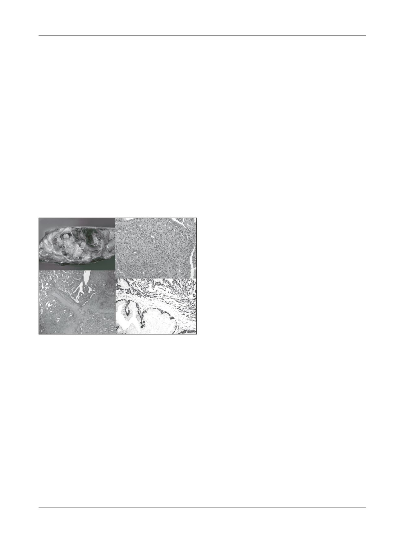
Figure 3
Pathologic findings. Macroscopic finding: (A) Gross
examination showed a large solid-cystic tumor. Microscopic findings:
(B) papillary neoplasia with sclerotic stroma (HE, 40x); (C) areas
containing carcinoma
in situ
(HE, 200x); (D) Immunohistochemistry
positive for myoepithelial cells (Calponin, 200x).
Mammary extensive papillomatous lesions represent
a clinical challenge, especially when observing a highly sus-
picious malignant tumor based on clinical and radiologi-
cal findings.
5
As core biopsy showed a benign lesion, an
open biopsy in the vegetative intracystic lesion was perfor-
med to improve material sampling. So, when a definitive
diagnosis of malignancy cannot be done because of discor-
dant findings, sampling limitations of a core biopsy
5
or
open biopsy, or limited sensibility of breast images,
5,6
re-
section of the entire lesion is mandatory
1-4,6
due to high as-
sociation with malignancy.
1,2,4
The open biopsy was an at-
tempt to improve the pathological results that were
hindered by limitation of diagnostic procedures and dis-
cordant findings. Also, the indication for diagnostic mas-
tectomy,
7
as seen in this case, is a fact that must be tho-
roughly discussed with the patient.
R
eferences
1.
Sydor MK, Wilson JD, Hijaz TA, Massey HD, Paredes ESS. Underestimation
of the presence of breast carcinoma in papillary lesions initially diagnosed
at core-needle biopsy. Radiology 242(1):58-62.
2.
Liberman L, Tornos C, Huzjan R, Bartella L, Morris EA, Dershaw DD. AJRIs
surgical excision warranted after benign, concordant diagnosis of papilloma
at percutaneous breast biopsy? ARJ. 2006; 186(5):1328-1334.
3.
Ueng SH, Mezzeti T, Tavassoli FA. Papillary neoplasms of the breast. Arch
Pathol Lab Med. 2009; 133(6):893-907.
4. Youk JH, Kin EK, Kwak JY, Son EJ. Atypical papilloma diagnosed by
sonographically guided 14-gauge core needle biopsy of breast mass. AJR.
2010;194(5):1397-492.
5.
LamWWM, Chu MCW, Tang APY, Tse G, Ma TKF. Role of radiologic features
in the management of papillary lesions of the breast. AJR 2006;186(5):1322-
1327.
6.
Eliada R, Chong H, Lylmarni S, Goldberg F,Muradai S. Papilllary lesions of
the breast: MRI, ultrasound, and mammographic appearances. AMJ J
Roentgenol.2012; 183(2): 264-71.
7.
Fenoglio C, Raffaele L. Sclerosing papillary proliferations in the female
breast. A benign lesion often mistaken for carcinoma. Cancer 1974; 33(3):
691-700.
A
C
B
D