


 22 / 116

V
ieira
RAC
et
al
.
518
R
ev
A
ssoc
M
ed
B
ras
2014; 60(6):518-519
IMAGE IN MEDICINE
Giant sclerosing papilloma mimicking locally advanced breast
carcinoma
P
apiloma
esclerosante
gigante mimetizando
carcinoma
de mama
localmente
avançado
R
ené
A
loisio
da
C
osta
V
ieira
1*
, S
ilvia
M
aria
P
rioli
de
S
ouza
S
abino
2
,
G
ustavo
Z
ucca
M
atthes
1
,
A
napaula
H
idemi
U
ema
W
atanabe
2
, L
ucas
F
aria
A
brahao
-M
achado
3
1
Department of Mastology and Reconstructive Surgery, Barretos Cancer Hospital – Pio XII Foundation, Barretos, SP.
2
Department of Radiology Breast Image Division, Barretos Cancer Hospital – Pio XII Foundation, Barretos, SP.
3
Department of Pathology, Barretos Cancer Hospital – Pio XII Foundation, Barretos, SP.
Study conducted at Barretos Cancer Hospital -- Pio XII Foundation, Barretos, SP
*Correspondence
Address: Rua Antenor Duarte Villela, 1331
Bairro Dr Paulo Prata
Postal Code: 14784 – 400
Barretos – SP
posgrad@hcancerbarretos.com.br http://dx.doi.org/10.1590/1806-9282.60.06.007Conflict of interest:
none
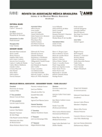
The patient was a 54-year-old Brazilian woman presen-
ting a progressive mass in the right breast. The clinical
exam showed a 9 x 8 cm tumor and a hardened axillary
lymph node. It was clinically considered a T3N1M0 breast
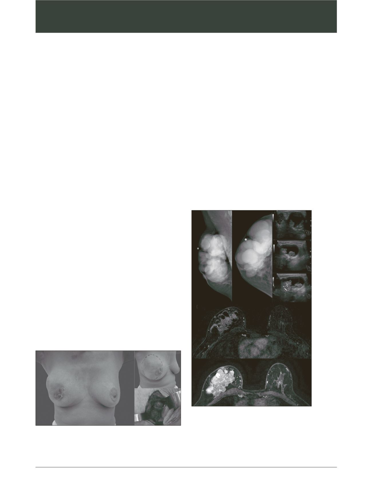
tumor (Figure 1A). Mammography showed multiple oval
formations, occupying the whole breast (Figure 2A). Ul-
trasonography showed the presence of multiple cysts,
many of which containing vegetating lesions with inten-
se vascular flow (Figure 2B) and absence of axillary lesion.
Magnetic resonance imaging showed multiple oval cysts
associated with vegetative lesions, a 4.7cm infiltrative area
near the pectoral muscle (Figure 2C), and normal enlar-
ged lymph node. As findings highly suspicious of malig-
nancy were noted, radiological staging was performed.
Abdominal ultrasound, bones scan and thoracic radio-
graphy showed absence of metastatic disease.
Figure 1
Clinical exam. (A) lump in right breast; (B) marked area
showing clinical localization; (C) open biopsy.
Figure 2
Radiologic findings. (A) Mammography: multiple
round image in the whole right breast; (B) breast ultrasound: cystic
mass with intense vascular flow; (C) MRI findings: infiltrative solid
mass with intense early enhancement and washout kinetic curve
associated with multiple cysts occupying the right breast.
A
A
B
B
C
C