


 14 / 139

S
ilva
PHRQ
da
et
al
.
486
R
ev
A
ssoc
M
ed
B
ras
2016; 62(6):485-489
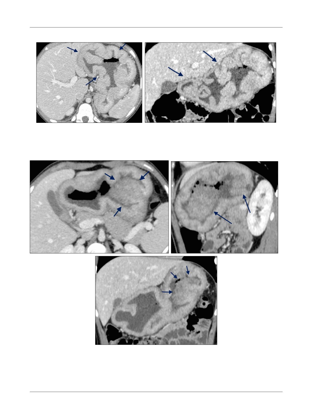
FIGURE 2
Axial (A), sagittal (B), and coronal (C) CT scans performed with contrast show two vegetating lesions (arrows) in the large gastric
curvature, in addition to diffuse parietal thickening.
A
B
C
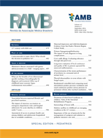
FIGURE 1
Axial (A) and coronal (B) CT scans performed with contrast show diffuse and redundant thickening gastric wall with no suspicious
lesions (arrows).
A
B