


 17 / 99

I
maging
features
of
idiopathic
granulomatous
mastitis
–
C
ase
report
R
ev
A
ssoc
M
ed
B
ras
2016; 62(4):303-306
305
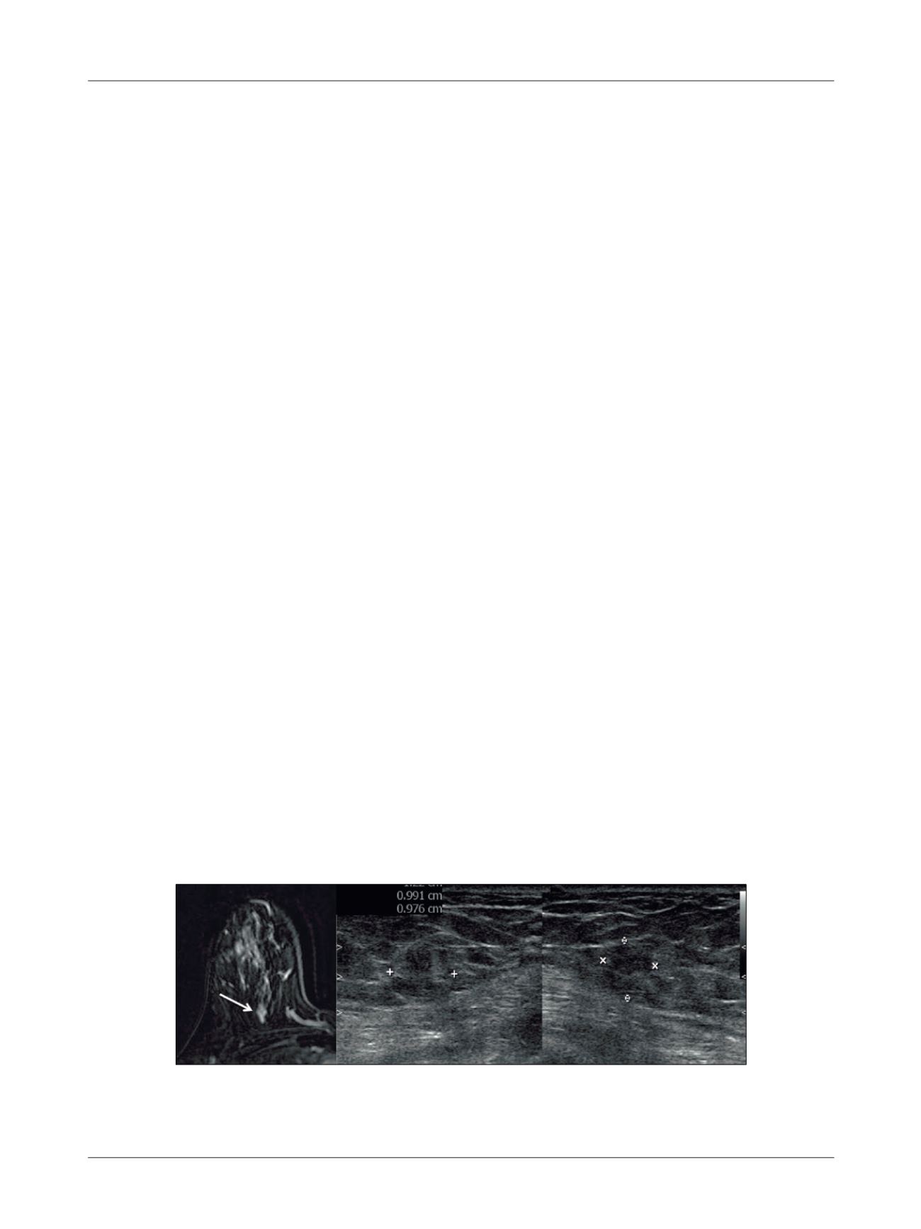
FIGURE 3
MRI (A) showing a mass enhancement at the upper quadrants of the right breast (arrow). Targeted ultrasound (B) revealing a
heterogeneous isoechoic mass in the same topographic of the MRI finding.
sity on T1 and T2 (Figure 2). MRI also revealed a mass
enhancement at the upper quadrants of the right breast,
which was subsequently identified in targeted ultrasound
(Figure 3).
The patient underwent ultrasound-guided percuta-
neous core needle biopsy on both breasts, which showed
marked chronic inflammation associated with granulo-
mas and microabscesses in breast lobules, more evident
to the left. No evidence of malignancy was found. Search
for etiological agents using Grocott, PAS and Ziehl-Neelsen
special staining yielded negative results. Thus, the final
pathological diagnosis was idiopathic granulomatous
mastitis.
D
iscussion
The case described above is typical of idiopathic granu-
lomatous mastitis in a patient with recent pregnancy his-
tory, who showed prolonged symptoms of a palpable mass
in the breast associated with inflammatory signs, and no
improvement after treatment with antibiotics. This diag-
nostic possibility should always be remembered in such
cases because the clinical and imaging findings can mim-
ic malignancy. Bilateral involvement is unusual and may
be associated with increased risk of recurrence and resis-
tance to medical treatment.
6
The most common mammographic finding is focal
asymmetry, not associated with distortions or microcal-
cifications; nodules with defined margins are less com-
mon. Sonographic findings include one or more irregular
hypoechoic masses associated with increased echogenici-
ty of the parenchyma without posterior acoustic shadow-
ing. Doppler study showed an increase of vascularization.
These imaging findings are nonspecific and mimic breast
carcinoma.
1,2,4
These findings on MRI are little known because there
are few reports in the literature. The most common is a
non-mass enhancement with the following distribution:
segmental, ductal and regional. The most common mass
findings are irregular margins with intense post-con-
trast peripheral enhancement. The kinetic curve is usu-
ally suspicious. However, the morphological findings of
lesions often do not distinguish them from malignant
lesions.
7
In the case above, MRI showed involvement of
the contralateral breast that had not initially been iden-
tified on conventional tests.
As described above, diagnostic confirmation can be
done by percutaneous core needle biopsy, which is more
accurate than fine-needle aspiration.
2,5
Microscopic anal-
ysis showed mixed inflammatory cell infiltrate (mononu-
clear and polymorphonuclear), abundant with histiocytes
within and outside of the breast lobules, non-caseating
granulomas and microabscesses (neutrophil cluster), and
absence of infectious etiologic agents.
2,3
Prognosis and treatment depend on the disease pre-
sentation. Steroid therapy is indicated for extensive cases,
but not for localized forms. Surgical removal is indicated
in refractory cases, but may be associated with the occur-
rence of fistulas and scars. Recurrence decreases when the
surgical margins are not compromised. The course of the
disease is usually long with a significant impact on qual-
ity of life. Prognosis is good; however, sometimes there is
delay in the improvement of the disease, and possible re-
currences and interventions can leave sequelae.
1,2,8
There-
fore, knowing this pathological entity, its clinical course
and imaging findings is important for proper medical
management, since this is a benign entity.
R
esumo
Aspectos de imagem na mastite granulomatosa idiopática
– Relato de caso
A mastite granulomatosa idiopática é uma afecção rara e
de etiologia desconhecida. Essa doença ocorre principal-
A
B
C