


 16 / 99

G
raziano
L
et
al
.
304
R
ev
A
ssoc
M
ed
B
ras
2016; 62(4):303-306
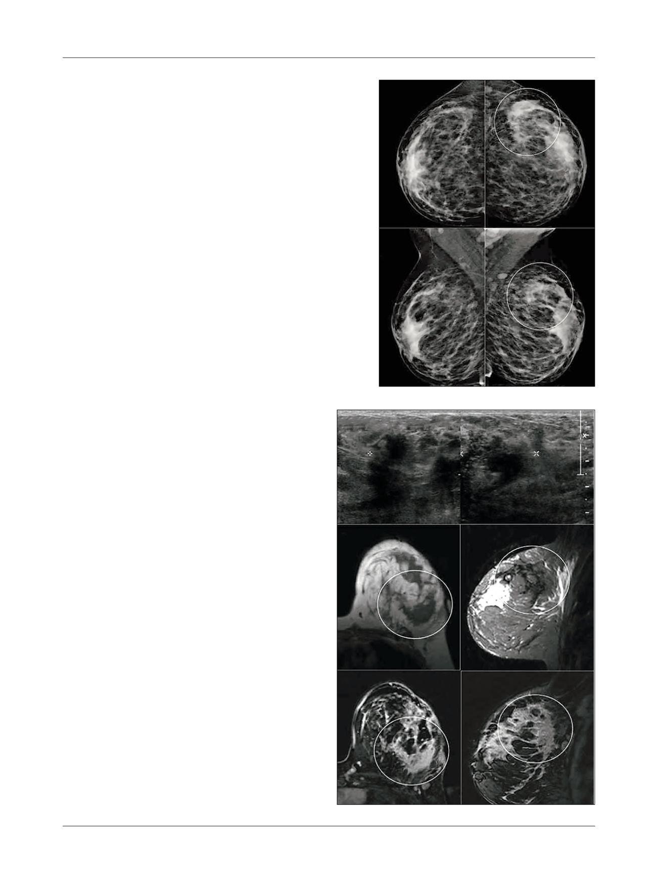
FIGURE 2
Ultrasonography showing a heterogeneous, predominan-
tly hypoechoic area, with indistinct margins, in the upper-outer quadrant
of the left breast (A and B). MRI showing an area of non-mass
enhancement with regional distribution occupying the lateral quadrants
of the left breast (highlighted), with low signal intensity on axial T1 (C)
and sagittal T2 (D) scans, presenting an heterogeneous internal
enhancement pattern (E – axial; F – sagittal).
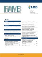
FIGURE 1
Mammogram revealing heterogeneously dense breasts, with focal
asymmetry in the upper-lateral quadrant of the left breast (highlighted).
A
B
C
D
E
F